DermaSensor Review: What the FDA Cleared AI Skin Cancer Device Actually Tells a Doctor
DermaSensor gives primary care physicians an objective signal about suspicious skin lesions in seconds. Its strongest result is sensitivity. Its hardest question is what happens after the device says a lesion needs further investigation.
Presented By Our Partners



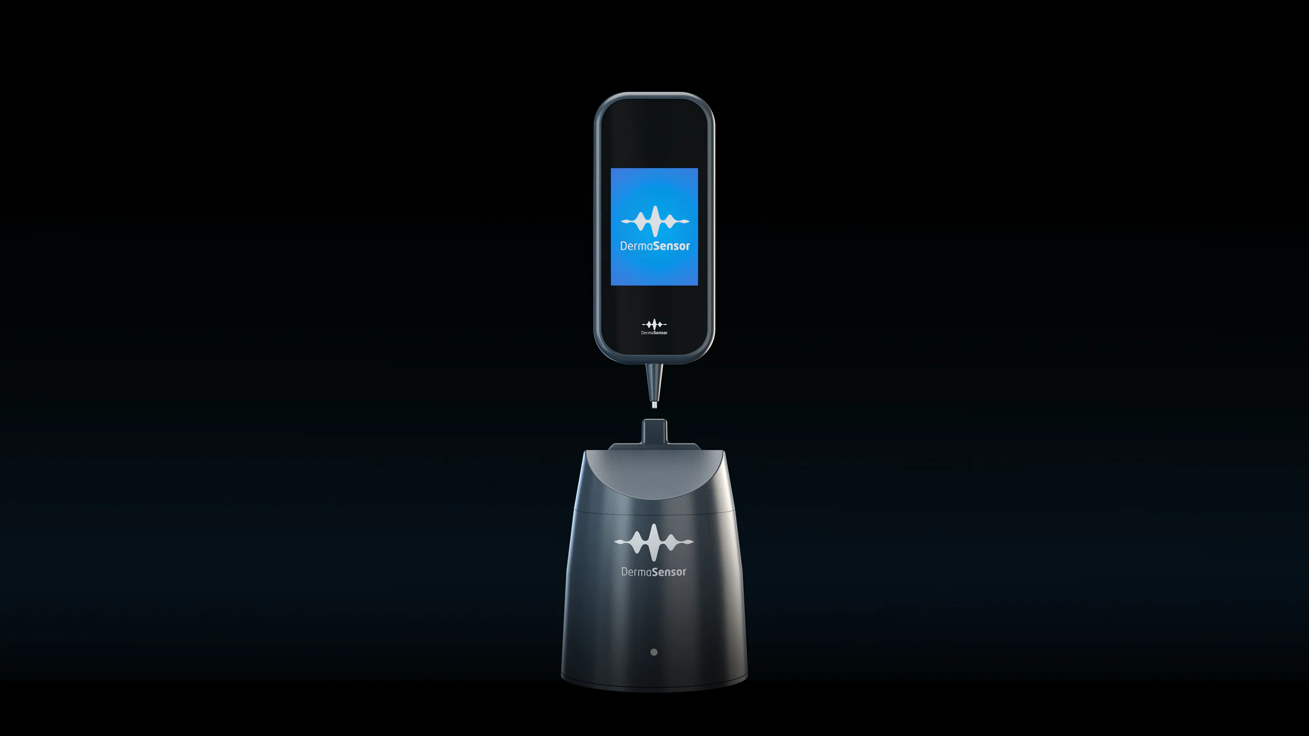
A suspicious mole creates an uncomfortable decision in primary care. Refer every uncertain lesion and dermatology queues grow longer. Reassure too quickly and a cancer may be missed. DermaSensor enters that decision as a handheld, prescription medical device that analyzes how light scatters beneath a lesion and returns one of two results: investigate further or monitor.
That is narrower than the phrase “AI skin cancer detection” suggests. DermaSensor does not diagnose cancer, replace a biopsy, screen healthy skin, or provide an at-home answer. The US Food and Drug Administration authorized it for non-dermatologist physicians evaluating lesions that are already suspicious in patients aged 40 and older. The result must be considered with the clinical examination.
The device is also a revealing case study in medical AI. Its pivotal evidence shows high sensitivity and low specificity. In plain English, it is designed to miss relatively few cancers, but it also sends many benign lesions toward further evaluation. That tradeoff may be reasonable when the disease is serious, but it belongs at the center of any honest review.
What Is DermaSensor and What Does a Scan Do?
DermaSensor is a wireless handheld device used by physicians who are not dermatologists. A physician places its disposable tip against a lesion that has already raised concern through history and visual examination. The device uses elastic scattering spectroscopy to sample optical properties associated with cellular and subcellular structure. A locked algorithm compares that signal with patterns learned from malignant and benign lesions.
The result is not a cancer type or pathology report. It is an adjunctive recommendation. “Investigate Further” supports referral or additional evaluation. “Monitor” supports continued clinical judgment rather than an automatic all-clear. The distinction matters because the FDA explicitly says the device is not a screening tool, cannot serve as the sole diagnostic criterion, and cannot confirm a skin cancer diagnosis.
DermaSensor targets melanoma, basal cell carcinoma, and squamous cell carcinoma. These are the three most common major skin cancers, but they do not carry equal risk. Basal cell carcinoma is common and usually slow growing. Squamous cell carcinoma can become invasive. Melanoma is less common but causes a disproportionate share of skin cancer deaths. A single combined sensitivity number can therefore conceal clinically important differences.
The practical appeal is speed. A primary care physician can obtain an objective signal during the visit instead of relying entirely on visual pattern recognition. That may be especially useful where dermatology access is slow or where a clinician sees skin lesions less frequently. It does not eliminate the specialist. It attempts to improve the decision about who should reach one.
DermaSensor Cost, Subscription, and Practice Access
DermaSensor is sold to medical practices as a monthly rental rather than a one-time device purchase. The company currently advertises a $199 monthly plan covering up to five patients and a $399 monthly unlimited plan. If a practice cancels, the device must be returned.
That pricing changes the buying calculation. A practice is not simply purchasing hardware. It is adopting a recurring clinical service whose value depends on patient volume, staff training, workflow fit, referral relationships, and reimbursement. At five patients per month, the limited plan represents about $40 per evaluated patient before any practice overhead. The unlimited tier becomes more economical as appropriate use rises, but only if the device is used on patients who match its authorized population and indication.
The rental model may reduce the initial capital barrier. It also creates ongoing dependence on the service. A practice evaluating the purchase should ask what is included in training and support, how disposable components are supplied, how results enter the medical record, what happens during downtime, and whether local payers reimburse the service. A low monthly sticker price can still produce poor value if the device sits in a drawer or creates referrals without a clear follow-up path.
For patients, this is not a product to order for home mole checks. Access runs through a participating medical practice. Cost to the patient may vary with the visit, the practice’s billing approach, and insurance coverage. A practice should explain any out-of-pocket charge before using the device.
FDA Authorization: What Was Actually Cleared?
The FDA granted DermaSensor De Novo authorization in January 2024 and classified it as a Class II prescription device. The regulatory category is a software-aided adjunctive diagnostic device for use by physicians on lesions suspicious for skin cancer. De Novo authorization establishes a new device type and special controls for future devices in that category. It is more precise to call DermaSensor FDA authorized or FDA cleared than FDA approved.
The exact boundaries are central to safe use. According to the FDA decision document, DermaSensor is indicated for lesions suggestive of melanoma, basal cell carcinoma, or squamous cell carcinoma in patients 40 and older. It assists a non-dermatologist physician in deciding whether to refer the patient to a dermatologist. It must be used with clinically relevant information, including visual analysis.
The device should not scan every mole as a population screening system. It should not substitute for dermatology or pathology. It should not turn a negative output into certainty. These are not cautious editorial additions. They are part of the authorized use.
The FDA also imposed post-market evidence requirements. The agency called for validation under anticipated real-world conditions and in demographic groups representative of the United States, including groups with limited melanoma representation in premarket studies and people with Fitzpatrick skin phototypes IV, V, and VI. That requirement makes demographic performance an active evidence question, not a footnote.
Accuracy: High Sensitivity Meets Low Specificity
In the pivotal evidence submitted for authorization, overall sensitivity was 95.5 percent, rising to 96.3 percent in patients aged 40 and older. Specificity was 20.7 percent. Negative predictive value was 96.6 percent, positive predictive value was 16.6 percent, and the reported number needed to biopsy was six.
Each number answers a different question. Sensitivity asks how often the device flags cancers among lesions that truly are cancer. Specificity asks how often it correctly returns a negative result among lesions that are benign. Positive predictive value asks how often an investigate-further result is actually cancer in the studied population. Negative predictive value asks how often a monitor result is benign in that population.
Featured Partner
Invest in the Infrastructure Behind Modern Medicine
As healthcare expands beyond hospital walls, the buildings and campuses supporting that shift are generating compelling returns for investors who move early. The Healthcare Real Estate Fund offers qualified investors direct access to a curated portfolio of medical office, outpatient, and specialty care facilities.
Learn More →
The high sensitivity is the strongest part of the case. A tool used to support cancer referral should place a premium on not missing malignancy. The low specificity is the price. Many benign lesions will receive a positive result, and a positive result does not mean a patient probably has cancer. With a PPV of 16.6 percent, most positive results in the study were not malignant.
Negative predictive value also needs context. A 96.6 percent NPV does not create a universal 96.6 percent guarantee for any mole. Predictive values change with disease prevalence and patient selection. The study involved lesions already considered suspicious enough for evaluation. Performance can shift when the device is used in a population with a different cancer rate or outside the authorized workflow.
This is why DermaSensor should be understood as referral support. It may add useful evidence to a difficult visual judgment. It does not collapse uncertainty into a diagnosis.
The Melanoma Gap Deserves Its Own Scrutiny
The combined cancer result can make performance look uniform. It was not. Reported sensitivity was 90.2 percent for melanoma, compared with 97.8 percent for basal cell carcinoma and 97.7 percent for squamous cell carcinoma.
Melanoma is the disease for which a false negative may carry the most serious consequences. A 90.2 percent sensitivity means the device identified most melanomas in the study, but not all. It also means a monitor result cannot overrule an alarming history, changing lesion, clinician concern, or established referral criteria.
The clinical utility study adds another tension. When physicians saw the device output, referral specificity fell from 44.2 percent to 32.4 percent, a statistically significant change with a reported p value of 0.026. The device helped reduce missed cancers, but it also made clinicians more likely to refer benign lesions. That is not necessarily a failure. Cancer triage often accepts more false positives to reduce false negatives. It is, however, a real downstream cost for patients and dermatology capacity.
An honest interpretation therefore holds three ideas at once. DermaSensor may improve sensitivity in primary care. Its melanoma sensitivity is lower than its performance for the other two cancers. Its output may increase unnecessary referrals. The best deployment would track all three, not celebrate only the first.
Where DermaSensor Could Help and Where Workflow Breaks
The clearest use case is a primary care practice whose clinicians encounter suspicious lesions but do not have dermatologist-level pattern recognition. The device supplies a standardized signal in a field where visual assessment can vary by training and experience.
Geography also matters. In communities with long dermatology waits, a more sensitive referral tool could help prioritize suspicious lesions and reduce casual reassurance based on appearance alone. It may also support conversations with patients who are uncertain about following through on a referral.
DermaSensor could be useful in practices serving older adults, since the authorized population begins at age 40 and skin cancer incidence rises with age. It may fit concierge medicine, multispecialty groups, urgent care, and rural primary care if staff understand the indication and have dependable dermatology referral pathways.
The wrong use case is indiscriminate scanning. Applying the device to low-risk lesions or younger patients would move beyond the studied and authorized population, lower positive predictive value, and potentially flood referral channels. A device designed to reduce missed cancers can become a referral amplifier when selection discipline disappears.
Limitations, Skin Tone, and Workflow Risks
Low specificity is the most visible limitation, but it is not the only one. Performance across diverse skin tones requires continued validation. The FDA specifically required post-market work that includes Fitzpatrick IV, V, and VI skin types and subgroups with limited melanoma representation. Until representative results are available, practices should avoid assuming identical performance across every population.
Workflow design determines whether an accurate signal becomes better care. A positive output has little value if the patient cannot obtain a dermatology appointment, the referral is not tracked, or biopsy results never return to the primary care record. Practices should define who communicates the result, who places and follows the referral, what happens after a monitor output when clinical concern remains, and how discordant cases are reviewed.
There is also automation bias. Clinicians may place too much confidence in a numerical or binary result because it feels objective. DermaSensor’s labeling directly resists that temptation by requiring the totality of clinical information. A concerning lesion remains concerning even when the device output is negative.
Finally, much of the accessible product narrative comes from the manufacturer. The pivotal and utility studies provide useful evidence, and independent analysis in npj Digital Medicine highlights the regulatory importance of the authorization. Long-term, real-world evidence should show whether deployment finds more consequential cancers, how many additional benign referrals and biopsies occur, whether performance is equitable, and whether follow-up is completed.
How DermaSensor Compares With Other Skin Evaluation Paths
A dermatologist’s examination remains broader than DermaSensor. Dermatologists combine lesion morphology, dermoscopy, patient history, whole-body context, and experience across many skin conditions. Biopsy with pathology remains the route to definitive diagnosis. DermaSensor sits before those steps.
Dermoscopy magnifies structures beneath the skin surface and can improve visual assessment in trained hands. It still depends heavily on expertise. DermaSensor instead produces an automated optical analysis, which may be more standardized for non-specialists but contains less contextual judgment.
Teledermatology can extend specialist access through photographs and remote review. Its strength is dermatologist interpretation. Its limitations include image quality, asynchronous workflow, and the possibility that a lesion requires in-person examination. DermaSensor provides an immediate office result but does not provide specialist judgment.
Consumer smartphone apps occupy a different category. They may encourage awareness, photography, or change tracking, but they are not substitutes for a prescription device used within a clinical assessment. Patients should not treat app output or DermaSensor marketing as permission to delay evaluation of a changing, bleeding, painful, or otherwise concerning lesion.
The most useful comparison is therefore not which tool “wins.” It is where each belongs: awareness at home, assessment in primary care, specialist examination in dermatology, and diagnosis in pathology.
What This Means for Your Health
DermaSensor is a promising addition to primary care because it addresses a real weakness: non-specialists must regularly decide whether a suspicious lesion deserves specialist attention. Its evidence supports a sensitive adjunctive signal, not autonomous diagnosis.
For a patient, the most important fact is simple. A DermaSensor result is one piece of a medical evaluation. An investigate-further result does not mean cancer has been diagnosed. A monitor result does not mean an alarming lesion can be ignored. Changes in size, shape, color, symptoms, or appearance still deserve professional evaluation.
For a practice, the decision is more complex. At $199 or $399 per month, the financial barrier is modest compared with many clinical devices. The operational burden lies downstream. Appropriate patient selection, informed interpretation, equitable performance monitoring, dermatology access, referral completion, and biopsy follow-up determine whether the technology improves care.
The verdict is cautiously favorable. DermaSensor appears most useful as a second signal for non-dermatologist physicians evaluating already suspicious lesions in adults 40 and older. Its high sensitivity is meaningful. Its low specificity, weaker melanoma sensitivity, demographic evidence gaps, and effect on benign referrals must remain visible. Medical AI earns trust when its tradeoffs are measured as carefully as its best number.
Frequently Asked Questions
How much does DermaSensor cost?
DermaSensor currently lists a $199 monthly rental for up to five patients and a $399 monthly unlimited plan. The device is returned if the practice cancels.
Can consumers buy DermaSensor?
No. It is a prescription medical device intended for use by non-dermatologist physicians, not an at-home mole scanner.
Does DermaSensor diagnose skin cancer?
No. It assists a physician’s referral decision for a lesion already considered suspicious. Diagnosis still requires appropriate specialist evaluation and, when indicated, biopsy and pathology.
How accurate is DermaSensor?
The pivotal evidence reported 95.5 percent overall sensitivity and 20.7 percent specificity. Sensitivity in patients 40 and older was 96.3 percent. Performance differed by cancer type, including 90.2 percent sensitivity for melanoma.
What does a negative DermaSensor result mean?
A monitor result lowers concern within the studied workflow but does not rule out cancer. The physician must still consider the lesion’s appearance, history, symptoms, and other clinical information.
Does DermaSensor work equally well across all skin tones?
That remains an important evidence question. The FDA required post-market validation in groups representative of the US population, including Fitzpatrick skin phototypes IV, V, and VI.
Is DermaSensor FDA approved?
The FDA granted De Novo authorization and classified DermaSensor as a Class II prescription device in 2024. Describing it as FDA authorized or FDA cleared is more precise than saying FDA approved.
Sources: FDA De Novo decision DEN230008; DermaSensor clinical resources; DermaSensor FDA authorization summary and study references; npj Digital Medicine regulatory and evidence analysis.


Similar Posts

Medicine Has Enough AI Pilots. It Needs a Power Grid.
Healthcare AI has enough pilots, alerts, and dashboards. The harder question is whether any of them close the loop from signal to ownership, action, completed care, and measured result.

From Prediction to Navigation: The AI Shift Healthcare Has Been Waiting For
Clinical AI has spent years predicting risk. The next test is harder: whether AI can help patients and clinicians choose the right next action and complete the path to care.
What “Clinically Validated” Actually Means
A single phrase covers at least four different evidentiary realities. Learning to tell them apart is the first move in reading healthcare AI.

How to Read a Healthcare AI Press Release
The healthcare AI press release is often the version of a claim most people ever encounter. This field guide shows how to read one in under ten minutes, using eight…
Who Decides Which AI Is Good Enough for Medicine?
Sixty-three separate scoreboards grade medical AI. Several are built or maintained by companies with a stake in the results, the scores cannot be compared to each other, and the most…

The New Race in Healthcare AI Is Not to Detect More. It Is to Finish the Job.
Healthcare AI is getting better at finding signals. The harder test is whether those signals turn into accountable action, completed care, and measured outcomes.